Peptide aggregation is the self-association of peptide molecules into higher-order structures that often reduces biological potency. It shows up as lost potency, hazy solutions, and assay results that don’t replicate. The fix starts before you ever open a vial: check the sequence for aggregation-prone stretches, control pH and concentration relative to the peptide’s isoelectric point, minimize surface exposure, and aliquot everything into single-use volumes the moment it’s reconstituted.
TL;DR:
- Aggregation-prone sequences, especially with aromatic or hydrophobic residues near the C-terminus, significantly increase the risk of peptide self-association.
- Surface interactions, pH near the peptide’s isoelectric point, and high concentrations accelerate aggregation, making buffer and storage conditions critical controls.
- The formation of mature fibrils is essentially irreversible, so early prevention via sequence modifications and careful handling is essential.
- Peptide purity and consistency from suppliers directly impact aggregation risk, with verified, high-quality starting material reducing unpredictable behaviors.
- Advanced predictive models now incorporate structural and contextual peptide information, enabling better pre-synthesis risk assessment.
Table of Contents
- What Is Peptide Aggregation and How Does It Form?
- Which Amino Acids Drive Peptide Aggregation the Most?
- Why Do Some Sequences Fail During Solid-Phase Synthesis?
- What Conditions Make Aggregation Worse?
- How Do You Actually Detect and Measure Peptide Aggregates?
- What Prevents Peptide Aggregation in Formulation and Handling?
- What’s the Right Way to Reconstitute and Store Peptides?
- How Supplier Quality Controls Reduce Aggregation Risk
- Do Post-Translational Modifications Change Aggregation Behavior?
- How Does Peptide Concentration Affect Nucleation Timing?
- Does Storage Time Affect Aggregation in Formulated Peptides?
- Can Peptide Aggregation Be Reversed Once It Happens?
- Can Machine Learning Actually Predict Aggregation Before You Synthesize?
- What the Research Actually Tells Us About Preventing Aggregation
- Where to Source Aggregation-Resistant Research Peptides
- Sources
What Is Peptide Aggregation and How Does It Form?
Peptide aggregation happens when individual peptide chains stop acting as independent molecules and start clumping into oligomers, amorphous precipitates, or ordered amyloid fibrils. The driving forces are the same ones that fold proteins in the first place, just pointed in the wrong direction: hydrophobic side chains seeking to escape water, backbone atoms forming hydrogen bonds into β-sheets, aromatic rings stacking against each other, and charged residues either attracting or repelling neighboring chains. Hydrophobic, hydrogen-bonding, and electrostatic forces all contribute, and which one dominates depends heavily on the peptide’s own sequence.
The process almost always follows the same basic arc: nucleation, then growth, then maturation. A small number of monomers first collide and form a nucleus, often the rate-limiting and most reversible step in the entire pathway. Once that nucleus exists, additional monomers add on rapidly, and the growing oligomer can become kinetically trapped in a low-energy conformation long before it reaches a true thermodynamic minimum. Molecular dynamics work has traced this in detail, showing that early oligomers frequently start disordered and only later reorganize into templated, parallel β-sheet structures that seed further growth, a pattern consistent with simulation studies of hierarchical aggregation pathways.
Surfaces deserve special attention because they punch above their weight. An air-liquid interface, a hydrophobic patch on a vial wall, or a nanoparticle contaminant can concentrate peptide locally and act as a nucleation template. Nanoparticle and surface-catalyzed studies show this can dramatically shorten the lag time before visible aggregation appears, sometimes by orders of magnitude compared to bulk solution alone.
The aggregates you end up with aren’t all the same animal:
- Soluble oligomers: small, often invisible to the eye, sometimes still partially active, and the hardest species to detect without dedicated tools.
- Amorphous aggregates: disordered clumps with no consistent internal structure, usually the product of rapid, uncontrolled precipitation.
- Amyloid fibrils: highly ordered, cross-β structures that are thermodynamically stable and resistant to most attempts at reversal.
Once fibrils form, they rarely go back. Mature fibrils sit in a deep kinetic trap; the same forces that built them hold them together against dilution, pH shifts, or mild denaturants. That’s why the field’s consensus, reflected across the physical stability literature on peptide therapeutics, treats nucleation prevention as the only strategy worth betting on.
Which Amino Acids Drive Peptide Aggregation the Most?
Not all sequences are equally risky, and the residues responsible are well cataloged at this point. Nonpolar, bulky side chains, think leucine, isoleucine, valine, phenylalanine, tryptophan, and methionine, minimize their contact with water by burying themselves against identical residues on neighboring chains. Aromatic residues carry extra risk because they add π-π stacking on top of the hydrophobic effect, and their position matters: work cataloging aggregation rules for short peptides found that aromatic residues sitting near the C-terminus disproportionately drive self-association compared to the same residue placed mid-chain.
On the other side of the ledger, charged residues (Asp, Glu, Lys, Arg) and polar uncharged residues (Ser, Thr, Gln, Asn) generally help keep peptides soluble and reduce aggregation by introducing electrostatic repulsion between molecules carrying the same net charge. This is why a single point substitution, swapping a leucine for a lysine at a critical position, can sometimes take a chronically difficult synthesis and make it tractable.
What’s changed the field more recently is the recognition that composition can matter more than order. Predictive work on tetrapeptides and pentapeptides shows aggregation propensity models can score short stretches with reasonable accuracy, and a separate line of research on amino acid composition in SPPS found that shuffling the order of residues while holding the overall composition constant often preserved the same aggregation behavior. That’s a genuinely useful, slightly counterintuitive result: two peptides with identical amino acid counts but different sequences can behave almost identically during synthesis, which means composition-vector screening can flag risk before you even settle on a final sequence.
Practical levers for dialing down risk include:
- Substituting a single hydrophobic residue for a charged or polar one at a flagged position.
- Adding a temporary solubilizing tag (a short charged stretch) that gets cleaved post-synthesis.
- Using protecting groups strategically to disrupt backbone hydrogen bonding during synthesis, not just to mask reactive side chains.
Pro Tip: Run your sequence through a composition-based aggregation propensity check before you commit to a synthesis route. If the score comes back high, look first at swapping a single aromatic or aliphatic residue near the C-terminus. That one change often does more than a full backbone-protection strategy.
Why Do Some Sequences Fail During Solid-Phase Synthesis?
Solid-phase peptide synthesis has its own aggregation problem, and it’s distinct from what happens in solution. As a chain grows on the resin, it can begin folding back on itself, forming resin-bound β-sheet structures that physically block the next incoming amino acid from reaching the reactive amine. Chemists call this a “difficult coupling,” and it’s usually not a coupling reagent problem at all. It’s aggregation happening in real time, on a solid support, mid-synthesis.
The tells are recognizable once you know what to look for: incomplete couplings that don’t improve no matter how much you increase reagent equivalents, a sudden drop-off in UV deprotection signal partway through a synthesis, and final crude purity that’s dramatically worse than a similar-length peptide with a different sequence. Since resin-bound aggregation and solution aggregation share root causes, composition-vector insights transfer directly here.
A few interventions consistently help:
- Lower the resin loading. Spreading chains further apart on the bead reduces the local concentration that drives interchain associations.
- Drop the coupling temperature slightly or extend reaction time rather than pushing more heat, which can accelerate aggregation instead of coupling.
- Insert pseudoproline dipeptides at positions prone to β-sheet formation. These temporarily kink the backbone and prevent the extended conformation aggregation needs.
- Use backbone-protecting groups (like Hmb or 2,4-dimethoxybenzyl) at specific amide bonds to physically block hydrogen bonding during the problematic stretch.
In-line UV monitoring during Fmoc deprotection gives you a live readout of coupling efficiency, and pairing that data with a composition-vector model of the sequence lets you predict, before synthesis even starts, which residues in the chain are most likely to cause a difficult stretch. That combination, real-time UV traces plus predictive composition screening, is quickly becoming standard practice in labs that run peptide synthesis routinely rather than occasionally.
Pro Tip: If a synthesis stalls at the same position across multiple batches, don’t just add more coupling reagent. Check whether that position sits inside a run of three or more hydrophobic or aromatic residues. If it does, a pseudoproline insertion two residues upstream usually fixes it faster than any reagent change.
What Conditions Make Aggregation Worse?
Sequence sets the baseline risk, but the environment decides whether that risk turns into an actual problem. The single most important variable is pH relative to the peptide’s isoelectric point (pI). Working within roughly 1 pH unit of the pI removes the net charge that normally keeps chains apart, and aggregation rates often spike right in that window. Choosing a buffer at least 1.5 to 2 pH units away from the pI, when the peptide’s intended use allows it, is one of the cheapest risk reductions available.
Concentration matters in a way that isn’t always intuitive. Every peptide has something like a critical aggregation concentration (CAC), a threshold below which nucleation is rare and above which it accelerates sharply. For many research peptides, a working stock in the 1 to 2 mg/mL range keeps you comfortably below that threshold while remaining practical for most assays; highly hydrophobic sequences may need to go lower still, sometimes paired with a co-solvent.
Beyond pH and concentration, a handful of physical stresses account for most of the aggregation events researchers actually encounter:
- Ionic strength can either shield electrostatic repulsion (encouraging aggregation) or disrupt it depending on the peptide’s charge profile, so it’s worth testing rather than assuming.
- Temperature swings, especially repeated warming to room temperature and back, give chains more kinetic energy to explore aggregation-prone conformations.
- Agitation and vortexing introduce air-liquid interfaces that act as nucleation surfaces, described in the same surface-catalysis mechanisms covered above.
- Freeze-thaw cycling causes cryoconcentration, where ice formation pushes peptide into a shrinking pocket of unfrozen liquid, spiking local concentration far above the bulk value.
- Container surfaces like glass and standard polypropylene can adsorb peptide and seed aggregation even when bulk concentration looks safe.
- Trace metal contamination can catalyze oxidation of methionine or cysteine residues, indirectly promoting aggregation; adding a chelator like EDTA at low concentration is a simple countermeasure.
Antioxidants such as methionine itself (as a sacrificial scavenger) or low concentrations of ascorbate can slow oxidative pathways in peptides that carry sensitive residues, though they need testing against your specific assay for interference.
How Do You Actually Detect and Measure Peptide Aggregates?
No single assay catches everything, which is the main reason aggregation gets missed in the first place. Soluble oligomers, amorphous clumps, and sub-visible particles all behave differently across analytical platforms, and a peptide can pass one test while failing another badly.
Size-exclusion chromatography (SEC) remains the workhorse for soluble aggregates because it separates by hydrodynamic size and gives a quantitative peak area you can track batch to batch. Its blind spot is real, though: SEC can under-report aggregates that stick to the column matrix, and its resolution starts to break down for very large or very small oligomeric species. Dynamic light scattering (DLS) and multi-angle light scattering (MALS) fill part of that gap, giving hydrodynamic radius and molar mass information without a column to interact with, though DLS is dominated by whatever large species are present and can miss a small population of dangerous oligomers hiding under a peak of monomer.
For sub-visible and visible particles, micro-flow imaging (MFI) or light obscuration (HIAC) methods count and size particles directly, and they map onto the particle-count thresholds referenced in USP <787> and <788> for injectable products, a useful anchor even for research-stage material headed toward eventual formulation work.
Structural confirmation comes from a different toolkit entirely. Thioflavin T (ThT) fluorescence and Congo red binding both report specifically on cross-β amyloid structure, giving you a fast, cheap readout of whether an aggregate is fibrillar rather than amorphous. Circular dichroism (CD) and FTIR spectroscopy both detect the shift toward β-sheet secondary structure that typically accompanies aggregation, and FTIR in particular is useful because it works even in turbid or concentrated samples where CD struggles. Mass spectrometry catches something none of the above can: covalent aggregates formed through disulfide scrambling or oxidative cross-linking, which behave completely differently from the physical, non-covalent aggregation that dominates most peptide instability.
The practical takeaway is orthogonality. Pairing SEC with either MFI or DLS catches both soluble and sub-visible populations, something neither method alone reliably does. A minimal defensible testing plan for most research peptides looks like SEC plus one structural method (ThT or CD) plus periodic visual/MFI inspection for particulates, escalating to MS if disulfide-containing residues are present.

What Prevents Peptide Aggregation in Formulation and Handling?
Prevention works in layers, and the most effective labs stack several of these rather than relying on one fix.
- Start with buffer and pH selection. Move at least 1.5 pH units away from the peptide’s isoelectric point when the application allows it, and choose an ionic strength that supports electrostatic repulsion between chains rather than screening it out.
- Add a non-ionic surfactant like polysorbate 20 or 80 at low concentration (typically 0.001% to 0.01%) to outcompete the peptide for surface adsorption sites at container walls and air-liquid interfaces. Go too high, though, and some surfactants introduce their own micelle-related assay interference, so titrate rather than guess.
- Use lyoprotectants such as trehalose or sucrose during freeze-drying, and freeze slowly and uniformly to avoid the localized cryoconcentration that seeds aggregation during the freezing step itself.
- Reach for co-solvents like DMSO or acetonitrile when a peptide’s hydrophobicity makes aqueous-only formulation impractical, but confirm compatibility with your downstream assay first. Some cell-based assays tolerate under 0.5% DMSO fine; others don’t tolerate it at all.
- When formulation alone can’t solve it, go back to the sequence. PEGylation, head-to-tail cyclization, or a cleavable solubilizing tag can all reduce the underlying aggregation propensity rather than just managing its symptoms downstream.
Low-bind plastic consumables deserve a specific mention here because they’re an easy, cheap fix that gets overlooked constantly. Surface-mediated aggregation research shows that standard polypropylene tubes and pipette tips can adsorb enough peptide at the surface to seed aggregation even when bulk concentration looks perfectly safe on paper. Switching to low-bind consumables, combined with a trace surfactant, closes off that entire pathway.
Pro Tip: If you’re troubleshooting a formulation that keeps failing SEC release testing, check surfactant concentration and container type before you touch the buffer chemistry. Adsorption-driven seeding is invisible until you specifically look for it, and it’s often the actual root cause behind what looks like a pH or ionic strength problem.
What’s the Right Way to Reconstitute and Store Peptides?
Reconstitution technique causes more aggregation than most researchers realize, and it’s entirely preventable with a consistent protocol.
- Choose the reconstitution solvent based on hydrophobicity, not habit. Highly hydrophobic sequences often need a small volume of DMSO or acetonitrile to fully dissolve before the aqueous buffer is added, rather than going straight into water and risking incomplete dissolution that looks fine but isn’t.
- Add solvent slowly, down the side of the vial, and swirl gently rather than vortexing. Vigorous agitation whips air into the solution and creates the interfacial nucleation surfaces covered earlier.
- Aliquot immediately into single-use volumes. This is the single highest-leverage habit in the entire prevention toolkit. Repeated freeze-thaw cycling causes cryoconcentration and generates fresh air-liquid interfaces every time a stock is thawed, and most researchers who trace an aggregation problem back to its source find a shared freeze-thawed stock sitting at the root of it.
- Decide filter versus centrifuge based on what you’re trying to remove. A 0.22 micron filter clears larger aggregates and particulates before an assay, but it can also strip out a meaningful fraction of active peptide if aggregation is heavy, so check post-filtration concentration rather than assuming recovery. Centrifugation followed by supernatant sampling is gentler when you need to preserve total protein content for a concentration readout.
- Verify concentration and activity after processing, not just before. A UV280 or BCA reading taken only from the original lyophilized vial tells you nothing about what survived reconstitution and filtration.
- Record every aggregation event, including turbidity observed by eye, unexpected SEC shoulders, or activity drop-off, with the batch number, buffer, and handling history attached. This turns a one-off frustration into a searchable dataset the next researcher on the project can actually use.
Pro Tip: Keep a simple reconstitution log for every peptide stock, noting solvent, volume, mixing method, and aliquot count. When a batch behaves unexpectedly six weeks later, that log is usually the fastest way to figure out whether the problem is the peptide or the handling.
How Supplier Quality Controls Reduce Aggregation Risk
A meaningful fraction of the aggregation problems researchers chase for weeks trace back to something that happened before the peptide ever reached the lab: inconsistent starting material. If a batch arrives with unknown truncation products, residual protecting groups, or variable purity from lot to lot, no amount of careful reconstitution technique will fully compensate.
Blankpeptides addresses that root cause directly. That verification matters more for aggregation work than it might first appear: a peptide with unresolved synthesis impurities or partial deletion sequences is far more prone to seeding aggregation than one confirmed clean, because those minor species often carry their own altered hydrophobicity and can act as nucleation triggers within an otherwise well-behaved sample.
A COA isn’t paperwork for its own sake. It’s the difference between troubleshooting your buffer chemistry and troubleshooting a contaminated starting material you never had reason to suspect.
Over 10,000 researchers have sourced peptides through Blankpeptides specifically because that verification removes one entire category of aggregation-related uncertainty from their experimental design. What that means in practice:
- Independent purity verification on every batch, not just periodic spot checks.
- COAs available per batch, so lot-to-lot variability is documented rather than assumed.
- USA-based manufacturing with consistent process control across production runs.
Starting clean doesn’t eliminate aggregation risk from pH, concentration, or handling. It does eliminate the possibility that your data is confounded by a problem baked into the vial before it arrived.
Do Post-Translational Modifications Change Aggregation Behavior?
Post-translational modifications (PTMs) don’t just add mass to a peptide, they can rewrite its aggregation profile entirely, sometimes in opposite directions depending on the modification and its position.
Phosphorylation typically adds negative charge at a specific site, which can suppress aggregation by boosting solubility and introducing electrostatic repulsion, particularly effective when the phosphorylation site sits near a hydrophobic stretch that would otherwise drive self-association. Glycosylation tends to work similarly, adding bulky, hydrophilic sugar groups that sterically block the close peptide-peptide contacts aggregation requires.
Other modifications push the opposite way. Deamidation of asparagine or glutamine residues can alter local charge and backbone flexibility in ways that sometimes increase aggregation propensity, especially in peptides already sitting close to their isoelectric point. Oxidation of methionine or tryptophan, often triggered by the trace metal contamination discussed earlier, changes side-chain polarity and can expose new hydrophobic patches that weren’t accessible in the unmodified peptide.
The practical implication is straightforward: a composition-based aggregation risk assessment done on the unmodified sequence can miss real risk if the peptide is going to carry a PTM in its final form. Any aggregation propensity screening should account for the modified residue’s actual chemical properties, not just the parent amino acid’s textbook classification.
How Does Peptide Concentration Affect Nucleation Timing?
Nucleation is inherently concentration-dependent, and that dependence isn’t linear. Below a peptide’s critical aggregation concentration, monomer-monomer collisions are rare enough that a stable nucleus essentially never forms within a practical timeframe. Cross that threshold, even modestly, and nucleation rates can increase sharply because collision frequency scales with concentration while the energetic cost of forming a nucleus stays roughly fixed.
This creates a lag phase, the period after a peptide solution is prepared but before any aggregate is detectable, that shortens dramatically as concentration rises. A peptide that looks perfectly stable at 0.5 mg/mL over a week might show measurable aggregation within hours at 5 mg/mL. Researchers who scale up an assay’s peptide concentration without re-testing stability at the new concentration are a common source of unexplained batch-to-batch inconsistency.
Characterizing the nucleation phase specifically, rather than just checking endpoint aggregation after some fixed incubation time, gives a far better picture of a peptide’s real risk profile. Time-course SEC or ThT fluorescence measurements taken at several time points can reveal a lag phase that a single endpoint measurement would completely miss, and that lag-phase data is exactly what tells you how much margin you actually have at your working concentration before nucleation becomes a practical concern.
Does Storage Time Affect Aggregation in Formulated Peptides?
Long-term stability and aggregation risk are tightly linked, and the relationship isn’t always obvious from short-term testing. A peptide formulation that passes release testing at time zero can still develop aggregates over weeks or months of storage, particularly if trace-level nucleation events that started during initial processing had time to slowly propagate.
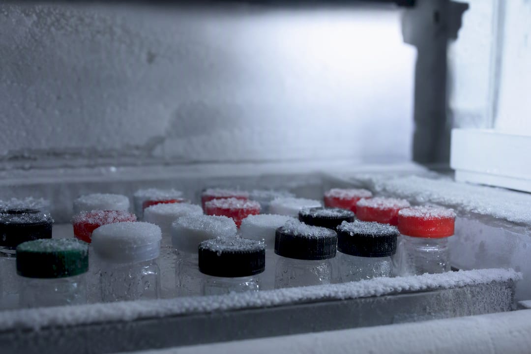
Temperature is the dominant variable. Storage at minus 20°C or minus 80°C dramatically slows the molecular motion needed for nucleation and growth, but it introduces its own risk if repeated freeze-thaw cycling happens during sampling. Refrigerated storage at 2 to 8°C is more convenient for frequently accessed stocks but gives aggregation pathways meaningfully more time to progress compared to deep-frozen storage, so the right choice depends on how often a given aliquot will actually be touched.
Lyophilized peptides generally show better long-term stability than liquid formulations because removing water removes much of the molecular mobility aggregation depends on, but reconstitution introduces its own risk window, one more reason the reconstitution protocol above matters as much as the storage condition itself. A formulation stability program worth trusting includes periodic SEC or ThT testing at defined intervals, not just an assumption that whatever passed release testing will remain stable indefinitely. Real-time stability data at the intended storage temperature, backed up by accelerated stability testing at elevated temperature, gives the clearest picture of how a given formulation will actually behave over its intended shelf life.
Can Peptide Aggregation Be Reversed Once It Happens?
The honest answer is: sometimes, but don’t count on it. Early-stage soluble oligomers, especially ones held together primarily by weaker, non-covalent interactions, can occasionally be partially disassembled by shifting pH away from the peptide’s isoelectric point, increasing ionic strength, or introducing mild detergents that disrupt the hydrophobic contacts holding the oligomer together.
Mature amyloid fibrils are a different story entirely. Once a peptide has organized into cross-β fibrillar structure, the aggregate sits in a deep kinetic trap that ordinary formulation adjustments rarely escape. Attempts to reverse mature fibrils typically require harsh conditions, strong denaturants, extreme pH, or elevated temperature, that themselves risk degrading the peptide or introducing new chemical modifications, meaning even a “successful” reversal often produces material that’s no longer identical to the original monomer.
This asymmetry is exactly why the field’s operating principle is prevention over correction. If you catch a formulation showing early SEC shoulders or a rising ThT signal, that’s the window to intervene, adjust buffer, reduce concentration, add a stabilizer, before the aggregate matures into something structurally locked in. Once fibrils are confirmed by ThT or Congo red binding, the more realistic plan is discarding that batch and adjusting the process upstream, rather than investing time trying to recover material that’s unlikely to return to a usable, well-characterized monomeric state.
Can Machine Learning Actually Predict Aggregation Before You Synthesize?
Composition-vector models were a genuine step forward, but the field has moved past treating amino acid composition as the only useful predictive signal. Newer approaches incorporate three-dimensional structural predictions, physicochemical property embeddings, and sequence-context features that composition-only models miss entirely.
Transformer-based architectures, adapted from the same family of models used in natural language processing, now treat peptide sequences the way language models treat sentences, learning which residue patterns and local contexts correlate with aggregation across large training sets of experimentally characterized peptides. This picks up positional and contextual effects, such as the C-terminal aromatic sensitivity noted earlier, that a pure composition vector, blind to order, structurally cannot capture on its own.
Some of the most useful recent work combines predicted secondary structure propensity (how likely a stretch is to favor β-sheet formation) with composition and sequence-context features simultaneously, producing a more complete risk score than any single input alone would give. These hybrid models still lean heavily on the core insight that composition is a powerful predictor in SPPS contexts, but they layer sequence-specific detail on top rather than discarding it.
For a working lab, the practical value isn’t in running your own transformer model. It’s in recognizing that aggregation prediction has become genuinely quantitative rather than purely empirical, and that a risk score generated before synthesis can meaningfully change which sequence variant you commit resin and reagents to first.
What the Research Actually Tells Us About Preventing Aggregation
The conventional advice on peptide aggregation still leans too hard on sequence order, treating it as the primary design variable when the composition-vector research increasingly says otherwise. If two sequences with the same amino acid inventory behave similarly under synthesis stress, then obsessing over exact residue placement while ignoring overall composition is solving the wrong half of the problem. That’s a real shift in how design decisions should get made, not a minor academic footnote.
What gets underrated constantly is handling discipline. Elegant formulation science means little if a stock gets vortexed, freeze-thawed four times, and stored in a standard polypropylene tube that’s quietly adsorbing peptide off the top of the solution. The cheapest, highest-leverage interventions, aliquoting, gentle mixing, low-bind consumables, get skipped in favor of chasing exotic excipients.
If you take one thing from all of this: screen composition before you synthesize, and control handling before you blame the formulation. Everything else is refinement on top of those two decisions.
— Blank Research Team
Where to Source Aggregation-Resistant Research Peptides
Clean starting material won’t fix a bad buffer choice, but it removes an entire category of confounding variables before your experiment even begins. Blankpeptides ships peptides finished in USA cGMP and FDA-registered facilities, with independent purity verification above 99% and a batch-specific COA behind every order, so the aggregation troubleshooting you do afterward is actually about your conditions, not an unverified vial.
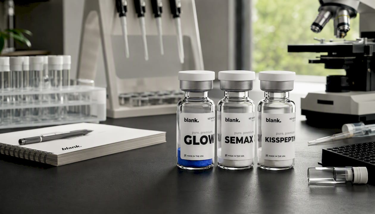
The current catalog includes research staples researchers are already building aggregation and stability studies around. GLOW is a frequently requested compound for metabolic and cosmetic-adjacent research applications. SEMAX sees steady demand in neuroscience-focused labs working on cognitive and neuroprotective pathways. KISSPEPTIN supports reproductive endocrinology research where receptor-binding fidelity depends heavily on maintaining a well-characterized, aggregate-free monomer population. Each product page carries batch-specific COA access, so you can confirm purity before it ever touches your buffer system.
Placing an order takes a few minutes through the Blankpeptides online catalog, and the technical support team is available to help match a compound and purity grade to your specific experimental design before you check out.